Academic Journal
Q1Radiology
About Radiology
Radiology is a scholarly journal published by Radiological Society of North America Inc.. SCImago 2025 places it in Q1 with an SJR of 4.388 and an H-index of 357.
Its listed coverage is 1931, 1936, 1945-2026 and its research categories include Radiology, Nuclear Medicine and Imaging (Q1). The 2025 dataset reports 540 documents and 14155 citations across the latest three-year reporting window.
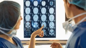
Radiology is a vital branch of medicine that utilizes advanced imaging technology to diagnose and treat diseases. From X-rays to MRI scans, radiology plays a crucial role in providing doctors with the images they need to make accurate diagnoses and plan treatments. This comprehensive guide will explore what radiology is, the types of imaging technologies used, and how it benefits healthcare.
What is Radiology?
Radiology refers to the medical specialty that employs imaging techniques to examine the inside of the body. The primary goal is to diagnose, monitor, and treat diseases and conditions by creating visual representations of the organs and structures within the body. These images help healthcare providers identify abnormalities, track disease progression, and plan surgeries or other treatments.
Radiology is divided into two main categories:
-
Diagnostic Radiology: Focused on diagnosing diseases through imaging.
-
Interventional Radiology: Involves using imaging techniques to guide minimally invasive procedures such as biopsies, catheter placements, and stent insertions.
Types of Radiology Imaging
Radiology encompasses a variety of imaging techniques, each with specific uses and benefits:
-
X-Ray: One of the most commonly used imaging techniques, X-rays use radiation to create images of bones and soft tissues. They are often used to detect fractures, infections, and tumors.
-
Computed Tomography (CT) Scan: CT scans combine X-ray images from different angles to create detailed cross-sectional images of the body. These are particularly useful for detecting cancers, internal injuries, and diseases affecting organs like the brain, lungs, and abdomen.
-
Magnetic Resonance Imaging (MRI): MRI uses powerful magnets and radio waves to generate detailed images of soft tissues. It’s commonly used to examine the brain, spinal cord, muscles, and joints. MRIs provide clearer images of soft tissues than X-rays or CT scans.
-
Ultrasound: Ultrasound imaging uses high-frequency sound waves to create images of internal organs and structures. It is safe, non-invasive, and commonly used in obstetrics to monitor pregnancies, as well as in cardiology, to examine the heart.
-
Nuclear Medicine: Involves the use of small amounts of radioactive substances to create images of organs and tissues. It’s commonly used for assessing the function of organs like the thyroid, heart, and bones.
-
Mammography: A specialized X-ray imaging technique used for early detection of breast cancer. Regular mammograms can help in detecting tumors before they become palpable.
Benefits of Radiology in Healthcare
Radiology offers several advantages in modern medicine:
-
Accurate Diagnosis: Radiological imaging helps doctors pinpoint the exact cause of symptoms and diagnose conditions early. This is essential for effective treatment and better patient outcomes.
-
Non-Invasive: Many radiology procedures are non-invasive, meaning no surgery or physical entry into the body is required. This reduces the risks associated with traditional surgical methods.
-
Early Detection: Regular imaging scans can detect health issues before they show symptoms. This is especially important in the early detection of cancer, heart disease, and neurological conditions.
-
Guided Treatments: Interventional radiology allows for minimally invasive procedures, such as removing a tumor or draining fluid, all while minimizing the need for open surgery.
Future of Radiology
Advances in technology continue to push the boundaries of what radiology can achieve. The future of radiology is leaning heavily on artificial intelligence (AI) and machine learning, which promise to improve image analysis, speed up diagnosis, and enhance treatment plans. AI can assist radiologists in identifying patterns in medical images, potentially leading to quicker, more accurate diagnoses.
Journal Metrics
Metrics can change by reporting year. Verify time-sensitive values with the publisher or indexing service.
Aims & Scope
Radiology is a critical field in modern medicine that plays a pivotal role in diagnosing and treating various health conditions. By utilizing imaging technologies such as X-rays, MRIs, CT scans, and ultrasounds, radiologists help physicians and specialists make accurate diagnoses, plan treatments, and monitor the progression of diseases. As medical technology continues to evolve, the scope of radiology expands, making it one of the most dynamic and essential branches of healthcare.
What is Radiology?
Radiology is the medical specialty that uses imaging techniques to view the internal structures of the body. These imaging methods allow healthcare professionals to detect and diagnose conditions such as cancer, fractures, infections, cardiovascular diseases, and neurological disorders. Radiology is divided into diagnostic radiology, which involves identifying diseases, and interventional radiology, which uses imaging guidance to perform minimally invasive procedures.
Growing Demand for Radiology
The demand for radiology services is rapidly increasing, driven by several factors. As populations age, the prevalence of chronic diseases such as heart disease, diabetes, and cancer rises, leading to an increased need for diagnostic imaging. Additionally, advancements in imaging technology have made procedures faster, more accurate, and less invasive, further driving demand. The rise of personalized medicine, where treatments are tailored to individual patients based on imaging data, is also contributing to the expansion of radiology services.
Advancements in Radiology
Technological advancements have significantly expanded the scope of radiology. Some of the most notable innovations include:
-
Artificial Intelligence (AI): AI is revolutionizing the field of radiology by improving image analysis, reducing human error, and speeding up diagnoses. AI algorithms can quickly detect abnormalities, such as tumors or fractures, that may be missed by human eyes, thus enhancing the accuracy of results.
-
3D Imaging: Advances in 3D imaging technology have allowed for more detailed and accurate images of the body's internal structures. This has proven particularly useful in planning complex surgeries, treating cancer, and monitoring treatment responses.
-
Nuclear Medicine: Nuclear medicine combines radiology with the use of radioactive substances to diagnose and treat diseases. This specialty is essential in detecting conditions such as cancer, thyroid disorders, and heart diseases.
-
Minimally Invasive Procedures: Interventional radiology has evolved to include a variety of procedures that are less invasive than traditional surgery. These procedures are often performed with the help of imaging technology, such as CT or ultrasound, and involve using small incisions or natural body openings.
Career Opportunities in Radiology
The scope of radiology also extends to numerous career opportunities. Radiologists, who are medical doctors specializing in interpreting medical images, are in high demand worldwide. Additionally, radiologic technologists and technicians who operate imaging equipment and assist in diagnosing diseases also play a crucial role. With the integration of new technologies, there is an increasing need for professionals with specialized skills in areas such as AI and medical imaging software development.
Recent Research Articles
Latest publications matched automatically by ISSN.
Exploring the Diagnostic Utility of Ferumoxytol for Brachial Plexus MR Neurography
Falko Ensle, Ek T. Tan, Sophie C. Queler, Yenpo Lin et al.
2026-09-01 · DOI: 10.1148/radiol.254051Ketamine Plus Midazolam versus Fentanyl Plus Midazolam for Sedation and Analgesia during Image-guided Procedures in Interventional Radiology: Randomized Clinical Trial
Amy R. Deipolyi, Maanasa Bommineni, Ashraf Ahmad, Adam Belcher et al.
2026-09-01 · DOI: 10.1148/radiol.260671Impact of Commercial Artificial Intelligence on Radiologist Reading Time for Pulmonary Nodule Evaluation at Chest CT
Jasika Paramasamy, Arlette E. Odink, Ties A. Mulders, Daniel Bos et al.
2026-09-01 · DOI: 10.1148/radiol.260484Convergence of Local and Systemic Therapies: A Comprehensive Society of Interventional Oncology Framework for 90 Y Radioembolization with Immunotherapy in HCC
Rony Avritscher, Edward Kim, Valerie Chew, Ghassan K. Abou-Alfa et al.
2026-09-01 · DOI: 10.1148/radiol.251415Black-White Racial Differences at Breast MRI
Pascal A. T. Baltzer, Paola Clauser
2026-09-01 · DOI: 10.1148/radiol.260622Histotripsy for Hepatocellular Carcinoma: From Bench to Bedside
Susu Luo, Xingyu Wei, Xinyu Lu, Zhenguang Wang et al.
2026-09-01 · DOI: 10.1148/radiol.254050CT Interpretation in the Artificial Intelligence Age: Turning Efficiency into Better Patient Care
Tae Iwasawa
2026-09-01 · DOI: 10.1148/radiol.262248Improving Recognition Performance and Reader Efficiency for Femoral-Neck Fracture Detection on Pelvic or Hip Radiographs Using Artificial Intelligence
Xin-Xiao Lin, Yu-Rui Qian, Shi-Hao Zhou, Han-Bin Shen et al.
2026-09-01 · DOI: 10.1148/radiol.253931Deceptive Eloquence in AI-assisted Thoracic Radiology
Alan B. McMillan
2026-09-01 · DOI: 10.1148/radiol.262291Styloid Process Elongation Leads to Carotid Artery Dissection in a Patient with Ankylosing Spondylitis
Jibao Wu, Yue Zhang
2026-09-01 · DOI: 10.1148/radiol.261271Bringing Inflammation’s Microvasculature into View: Super-Resolution US in Crohn Disease Activity—Early Career Perspective
Shailendra Katwal, Victoria Chernyak
2026-09-01 · DOI: 10.1148/radiol.262452Falling Fruit Sign: Pelvic Splenosis Supplied by the Celiac Trunk
Ji Zhang, Jiaji Liu
2026-09-01 · DOI: 10.1148/radiol.261804Beyond Burnout in Radiology: A Call to Action in the AI Era
Phillip M. Boiselle
2026-09-01 · DOI: 10.1148/radiol.261218Performance of Photon-counting CT for Assessing Pretreatment Breast Cancer: Comparison with Mammography, MRI, and 18 F-FDG PET/CT
Qiuyi Fu, Yufei Zeng, Rui Chang, Zhihan Xu et al.
2026-09-01 · DOI: 10.1148/radiol.253531A Progressive Shift in Procedural Sedation: Is It Time to Embrace Ketamine in Interventional Radiology?
Clifford R. Weiss, Lauren S. Park
2026-09-01 · DOI: 10.1148/radiol.262019Brain MRI Assessment of Anterior Inferior Cerebellar Artery Aneurysm Formation after Stereotactic Radiosurgery for Vestibular Schwannomas
Motoyuki Umekawa, Yuki Shinya, Hirotaka Hasegawa, Satoru Miyawaki et al.
2026-09-01 · DOI: 10.1148/radiol.261112Evaluation of Super-Resolution Contrast-enhanced US in Crohn Disease Activity Assessment
Saidaitiguli Aihemaiti, Manying Li, Ming Xu, Ren Mao et al.
2026-09-01 · DOI: 10.1148/radiol.253558Ethical Implications of the Invisibility of Intracellular Electrolytes
Juan Sebastián Rodríguez Sazipa
2026-09-01 · DOI: 10.1148/radiol.260782Sharpening the PET Amyloid Signal with MRI
Akshay S. Bedmutha
2026-09-01 · DOI: 10.1148/radiol.262286Closing the Gap in Femoral-Neck Fracture Detection: AI Assistance with Pelvic and Hip Radiographs
Dyan V. Flores, Tatiane Cantarelli
2026-09-01 · DOI: 10.1148/radiol.262353Reviews
Community Reviews
Version History
April 21, 2025 at 6:52 am
April 21, 2025